During any physical activity or movement, the human body interacts with the external environment and experiences external forces. These forces cause vibrations and oscillations within the soft tissues of the body. For example, during gait the legs alternate between the stance and swing phases. It is during heel strike of the stance phase that the gastrocnemius undergoes vibrations from the impact and must actively recruit muscle fibres to dampen the amplitude of these vibrations. The transmission of vibrations through the body is regulated by bone, cartilage, soft tissues, synovial fluids, joint kinematics and muscular activity (Cardinale & Wakeling, 2005). Joint kinematics and muscle activity, which can efficiently and effectively dampen vibration over a short time frame, are used by the body to change its vibrational response to external forces. Dr. Benno Nigg, at the University of Calgary, has implied that the body has a strategy of “tuning” its muscle activity to reduce its soft tissue vibrations in an attempt to reduce harmful effects (Nigg, 2001). According to this theory, the magnitude of the muscular response is related to the interaction between the amplitude and frequency of the vibration input and the intrinsic neuromuscular properties. It has also been suggested, and somewhat significantly shown in the research literature, that vibrations can be used in vibration therapy and as a training aid.
The Benefits of Vibromax Therapeutics Vibration Therapy
Some benefits of vibration training suggested in the literature include an increase in average force and power (Bosco et al., 1999), muscle blood flow (Kerschan-Schindl et al., 2001), proprioception (Fontana et al., 2005) and hormone secretions (Goto & Takamatsu, 2005). Other benefits, which have not been well studied in the literature but have been reported, are soft tissue and joint mobilizations, breakdown of scar tissue, and decrease in pain and muscle inhibition. The developers of VMTX have taken the benefits of vibration training and incorporated them in a therapeutic manner utilizing various myofascial kinetic chains.
Muscle Activation, Force and Power
It is well-known that primary afferent endings of motor spindles are particularly sensitive to vibration. It can be suggested that vibration stimuli cause excitation of these afferents and recruit more receptors which, in turn, activate a fraction of alpha-motoneurons whose discharges recruit previously inactive muscle fibers into contraction (Jordan et al., 2005). Unlike vibration therapy that is directly applied to a tendon or a specific muscle, the vibratory wave transmitted to a distal link propogate through proximally located muscle groups and activates an enormous number of vibratory sensitive muscle receptors. As a result, a large number of additional motor units can be involved in a motor response. Another explanation relates to the proprioceptive feedback of Ia afferents and stretch sensitivity of the muscles. When the muscle is pre-stretched and active prior to concentric contraction, the stretch reflex greatly contributes to the force development (Komi, 2000). It is known that force enhancement in stretch-shortening cycle exercises is strongly affected by reflexive facilitation of an efferents caused by Ia afferents (Ross et al., 2001). Similarly, dynamic vibration stimulation exercises are performed when the muscles are preliminary activated by vibratory waves causing Ia afferent inflow which produce excitatory effect on the alpha-motoneurons (Jordan et al.,2005). It has been suggested that the vibration stimuli facilitate stretch reflex, which can have an impact on force and power generation.
Muscle Blood Flow with Vibration Therapy
In an experiment conducted by Kerschan-Schindl et al., (2001), the authors showed a significant increase in muscle blood volume and in mean blood flow velocity after vibration therapy of only three minutes duration. This acute response was attributed to the effect of vibrations in also suggested that vibration may represent a mild form of exercise for the cardiovascular system (Kerschan-Schindl et al., 2001; Rittweger et al., 2002; Yamada et al., 2005).
Proprioception
There are many different kinds of receptors concerned with monitoring the body’s actions. Typically, when analyzing proprioception, four areas or senses have to be assessed:
1. Kinesthetic sense (the sense of positioning and movement)
2. Sense of tense
3. Sense of balance
4. Sense of effort or of heaviness
The sense of effort is usually thought to be centrally generated and does not require any feedback from peripheral receptors. It will, therefore, not be given any further attention. This brief review will focus on kinesthetic sense, and in particular, sense of static limb position. It is commonly recognized today that the muscle proprioceptive feedback generated by motor activities mainly contributes to the awareness of body position and movements (Gandevia & Burke, 1992; Roll et al., 2000). This current thinking is derived from the results of studies where muscle spindle afferent inputs were stimulated. In addition, it is clearly established that input arising from a single spindle ending has no perceptual relevance. Instead, inputs from an ensemble of muscle receptors seem to be required for a movement to be perceived. The research of Ribot-Ciscar (2003) has shown that the tendon excites the primary endings of the muscle spindles and induces reflex muscle contractions to help improve muscle function. It seems that vibration not only has an effect on the muscles and tendons to provide feedback but also on the joint structures. This means that additional sensory motor effects through the proprioceptive joint mechanoreceptors. The activation of joint mechanoreceptors and the stimulation of the gamma efferents sensitize the spindles and result in increased muscle stiffness and joint stability.
MYOFASCIAL KINETIC CHAINS: ACTIVE OR PASSIVE COMPONENTS?
Fascia can be defined as “dense irregular connective tissue sheets in the human body” (Schleip et al., 2005). Traditionally it was assumed that fascia acted as a passive structure that contributed to biomechanical behaviour of certain intertwined anatomical structures. More recent literature has shown, however, that fibroblasts, which are cells that give rise to fascial tissue, have the ability to express the gene for smooth muscle action (Spector, 2002). If triggered, these cells have the potential to generate contractile force and increase fascial stiffness (Bisson et al., 2004; Spector, 2002). In addition to opposing mechanical advantage, these cells can influence the mechanoreceptors and affect proprioceptive function. Recent literature has shown that the direct stimulation of a tissue may have a more dramatic effect than indirect vibratory stimulation. The direct vibratory stimulation of tissues, as found in VMTX, assures that the desired tissue is affected by the specific amplitude and frequency set by the practitioner. With indirect stimulation, the surrounding tissues will attenuate the vibrations before they reach the desired tissue. Thus, direct vibratory stimulation may facilitate a more efficient and effective therapeutic effect on the desired tissue. It is for this very reason that VMTX strives to treat the symptomatic tissue prior to treating the surrounding myofascial kinetic chains.
Vibromax Therapeutics Protocol
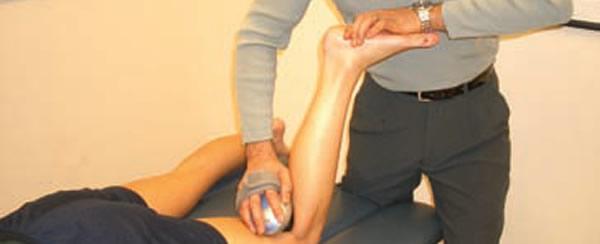
VMTX practitioners treat the specific injured tissue along with the myofascial kinetic chain where the symptomatic tissue may lie. For example, if a runner comes in with a strain of the medial head of gastrocnemius, the practitioner will first apply the VMTX hand-held apparatus to the injured fibres of the muscle for one minute. The vibration is then applied along the entire length of the gastrocnemius for an additional minute while the patient is actively lengthening and shortening the muscle. Then, vibration therapy is applied to the entire myofascial kinetic chain of which the gastrocnemius is a part. In this example, the corresponding kinetic chain is the superficial lower limb chain, which consists of the dorsal sacral ligament, sacrotuberous ligament, hamstrings, gastrocnemius, plantaris, soleus, Achilles tendon, the plantar fascia and plantar aponeurosis. Subsequently, the patient is instructed on stretching and strengthening exercises for that specific muscle and myofascial kinetic chain in order to prevent reoccurrence.
VIBRATION THERAPY CONCLUSION
The most encouraging aspect of this easy-to-learn technique is that we have clearly outlined the various anatomical structures with respect to their intimate anatomy, functional and clinical relevance, localization and palpation, and detailed treatment procedures. VMTX Vibromax Therapeutics soft tissue therapy is appropriate not only for chiropractors and chiropractic students, but all practitioners who are interested in treating soft tissue injuries, and enhancing their patients’ health, well-being and performance.
VIBRATION THERAPY CERTIFICATE PROGRAM
Dr. Kazemi, Doctor of Chiropractic, has developed a new 3 step certificate program for VMTX Vibromax Therapeutics Soft Tissue Therapy:
- Study the Treating Lower Limb Injuries eBook
- Take the online exam
- Attend the Lower Limb Soft Tissue Massage Therapy Workshop at the DoctorK Chiropractic Clinic Toronto
REFERENCES:
Nigg BM. (2001). The role of impact forces and foot pronation: A new paradigm. Clinical Journal of Sports Medicine, 11(1): 2-9.
Rittweger J, Ehrig J, Just K, Mutschelknauss M, Kirsch KA, Felsenberg D. (2002). Oxygen uptake in whole-body vibration exercise: influence of vibration frequency, amplitude, and external load. International Journal of Sports Medicine, 23(6): 428-32.
Ribot-Ciscar E, Bergenheim M, Roll JP, Albert FJP. (2003). Facilitation of triceps brachii muscle contraction by tendon vibration after chronic cervical spinal cord injury. Journal of Applied Physiology, 94(6): 2358-2367.
Roll JP, Bergenheim M, Ribot-Ciscar E. (2000). Proprioceptive population coding of two-dimensional limb movements in humans: II. Muscle- spindle feedback during “drawing-like” movements. Experimental Brain Research, 134(3): 311-21.
Ross A, Leveritt M, Riek S. (2001). Neural influences on sprint running: training adaptations and acute responses. Sports Medicine, 31(6): 409-25.
Schleip R, Klingler W, Lehmann-Horn F. (2005). Active fascial contractility: Fascia may be able to contract in a smooth muscle-like manner thereby influence musculoskeletal dynamics. Medical Hypotheses, 65: 273- 277.
Spector M. (2001). Musculoskeletal connective tissue cells with muscle: Expression of muscle actin in and contraction of fibroblasts, chondrocytes, and osteoblasts. Wound Repair and Regeneration, 9(1): 11-18.
Yamada E, Kusaka T, Miyamoto K, Tanaka S, Morita S, Tanaka S, Tsuji S, Mori S, Norimatsu H, Itoh S. (2005). Vastus lateralis oxygenation and blood volume measured by near-infrared spectroscopy during whole body vibration. Clinical Physiology & Functioning Imaging, 25(4): 203-8.
Bisson MA, Mudera V, McGrouther DA, Grobbelaar AO. (2004). The contractile properties and responses to tensional loading of Dupuytren’s Disease – derived fibroblasts are altered: a cause of the contracture? Plastic Reconstruction Surgery, 113(2): 611-621.
Bosco C, Colli R, Introini E, Cardinale M, Tsarpela O, Madella A, Tihanyi J, Viru, A. (1999). Adaptive responses of human skeletal muscle to vibration exposure. Clinical Physiology, 19(2):183-7.
Cardinale M, Wakeling J. (2005). Whole body vibration exercise: Are vibrations good for you? British Journal of Sports Medicine, 39(9): 585-589.
Fontana TL, Richardson CA, Stanton WR. (2005). The effect of weight-bearing exercise with low frequency, whole body vibration on lumbosacral proprioception: A pilot study on normal subjects. Australian Journal of Physiotherapy, 51(4): 259-263.
Gandevia SC, Burke D. (1992). Does the nervous system depend on kinesthetic information to control limb movement? The Behavioral and Brain Sciences,15: 614-632.
Goto K, Takamatsu K. (2005). Hormone and lipolytic responses to whole body vibration in young men. Japanese Journal of Physiology, 55(5): 279- 84.
Jordan MJ, Norris SR, Smith DJ, Herzog W. (2005). Vibration training: an overview of the area, training consequences, and future considerations. Journal of Strength & Conditioning Research, 19(2): 459-66.
Kerschan-Schindl K, Grampp S, Henk, C, Resch H., Preisinger E, Fialka-Moser, V, Imhof H. (2001). Whole-body vibration exercise leads to alterations in muscle blood volume. Clinical Physiology, 21(3): 377-382.
Komi, PV. (2000). Stretch-shortening cycle: A powerful model to study normal and fatigued muscle. Journal of Biomechanics, 33(10): 1197-1206.
Dr. Kazemi is a doctor of chiropractic that has been practising as a Toronto Chiropractor for over 20 years. He is a sports chiropractor providing chiropractic, acupuncture, sports medicine, rehabilitation, soft tissue therapy, and other chiropractic services. He has invented innovative chiropractic tools and educates practitioners with chiropractic seminars Toronto.


